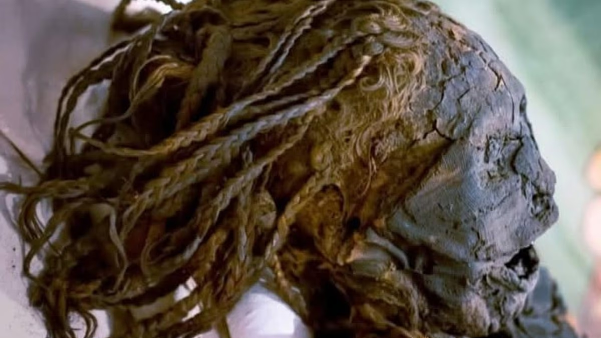

Scientists have uncovered new details about an ancient Egyptian woman known as the “Braided Lady” after digitally examining her mummy with high-resolution CT scans.
Researchers studied the remains, thought to date from between Egypt’s 18th and 21st Dynasties, looking for clues about the woman’s life, health and death.
Among her most striking features is her long, carefully braided hair, which has survived remarkably intact for more than 3,000 years.
Her teeth provided another important clue. They were severely worn and damaged, likely because of the gritty, sand-contaminated foods commonly eaten in ancient Egypt.
The scans also identified gum disease, a fractured molar and an incisor that had shifted backwards towards the throat.
Researchers say the unusual dental damage may be evidence that the woman was involved in an ancient funerary ceremony.
“Dental wear and pathology were observed, likely reflecting the abrasive diet of ancient Egyptians, and unique dental findings suggested participation in the ‘Opening of the Mouth’ ritual,” the team wrote in the journal Radiology Case Reports.
The ceremony was carried out after death and was intended to restore the deceased person’s senses for use in the afterlife.

Her long, intricately braided hair is one of the mummy’s most remarkable features and has remained intact for more than 3,000 years

High-resolution CT scans digitally unwrapped the mummy, revealing gum disease, a fractured molar and an incisor displaced towards the throat
The woman was first found in a cache of mummies in Egypt before being transported to Canada in 1860.
She is thought to have lived between 1077 BC and 1292 BC, around 3,000 to 3,300 years ago, when ancient Egyptian civilisation was at the height of its power.
A team led by a researcher at the University of Michigan used CT imaging to investigate the interior of her preserved remains.
The technique combines hundreds of X-ray images to produce detailed three-dimensional views, allowing researchers to “unwrap” a mummy digitally without damaging it.
The examination showed that her body was exceptionally well preserved, with both bone and soft tissue still visible.
The scans indicated that the embalmers had left some internal organs inside the body, including remnants of the liver.
Dried brain tissue was also present, suggesting that her brain had not been removed during mummification.
The researchers said this combination of preservation methods matches burial practices associated with wealthy non-royal Egyptians during the New Kingdom period.

Although the mummy was examined in detail, researchers found no fatal injury or clear disease that could explain her death

CT scans showed that some internal organs, including remnants of the liver, had been left inside the body during embalming (yellow arrow)
The scans further showed that the Braided Lady was a petite woman, measuring approximately 4ft 11in (151cm) in height. She died between the ages of 35 and 50.
Despite the extensive investigation, the team found no evidence of a fatal injury or obvious illness that could account for her death.
Small calcified deposits visible in the scans could indicate that she had suffered a long-term parasitic infection, according to the experts.
Such infections were common in ancient Egypt, where many people lived near the Nile and depended on its water for drinking, bathing and farming.
However, the evidence is inconclusive and does not indicate that the infection was fatal.
The cause of the Braided Lady’s death therefore remains unknown, even thousands of years after her burial.
“Despite limitations posed by tissue desiccation and lack of historical records, this multidisciplinary radiological assessment advances our understanding of ancient Egyptian funerary practices and demonstrates the value of modern non-invasive imaging in reconstructing the lives and deaths of archaeological individuals,” the team concluded.


